Corbin Jensen

Background & Contact Information
Position (2022 – Present)
Education: PhD in Cancer Biology (2022) at the University of Arizona (Advisor: Noel Warfel)
Fellowships & Awards: NIH F31 Ruth L. Kirschstein National Research Service Award (2020-2022), AACR Scholar in Training Award (2022), NIH T32 Integrative Cancer Scholar Trainee Award (2020), Jim Cockrum Innovation Award (2019)
Email: jensencc@unc.edu
Research Information
Before joining the Peifer lab my research explored the topics of tissue differentiation and cell migration. During my undergraduate research I studied two key pathways that facilitated the transition of prostate cells from a monolayer of basal cells into a differentiated bi-layer of basal and luminal cells. Defects and errors in this process represent a potential primary step in the oncogenic process of cancer formation. My PhD research shifted my focus into the realm of cancer biology. Specifically, I studied how tumor cells leave their primary location, invade into surround tissues and ultimately metastasize to distant organs within the body. This resulted in a deep dive into the underlying mechanisms responsible for cell motility, migration and invasion; namely how changes in actin dynamics were regulated by changes in oxygen levels to drive cell migration and invasion. My work here in the Peifer lab builds on both of these areas in unique and interesting ways as embryonic development encompasses both differentiation and cell motility with many more fascinating processes involved.
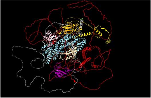
Understanding the mechanisms that link cell-cell adherens junctions (AJs) to the actin cytoskeleton is essential. During embryonic development the interactions between AJs and the cytoskeleton are both dynamic, as the embryo and its component cells are constantly changing shape, as well as robust since the cytoskeleton generates a high level of pulling force on cell-cell contacts as the cells change shape and move. Disrupting interactions between AJs and the cytoskeleton disrupts diverse morphogenetic events, from gastrulation to germband extension to dorsal closure. Over the last 15 years, joint efforts in the field revealed that the connection between AJs and the cytoskeleton involves a complex network of proteins and not a simple linear pathway as once thought. A central member of this network, and a superb entry point to better understanding these complex connections, is the Drosophila protein Canoe. Canoe and the mammalian homolog Afadin contain multiple domains, some of which have known binding partners and are well studied, e.g. Rap1-binding RA domains and the E-cadherin-binding PDZ binding domains. However, Canoe and Afadin also contain a large C-terminal region that is predicted to be intrinsically disordered (IDR), comprising over 1/3 of the total protein. The IDRs of both Canoe and Afadin are quite different in length, charge and amino acids composition, and have little sequence conservation. Several of the regions in the IDR that are most conserved over evolution are predicted to be alpha-helical by AlphaFold. One of these helical regions corresponds to a known F-actin-binding site. Combining bioinformatic, genetic and cell biological analyses, we seek to define the function of the IDR on Canoe function and localization during embryonic development. In Silico data suggests that both the IDR and the FAB domain of Canoe are capable of binding actin either in coordination with or independently of each other. We have generated mutants that completely remove the IDR from Canoe in vivo. Our preliminary results reveal that loss of the IDR in Canoe dramatically reduces function and largely, but not completely, eliminates localization of Canoe to cell junctions. Further in-depth analysis of the IDR will provide a clearer picture of the mechanisms the drive both embryonic development as well as the interaction between junctions and the cytoskeleton.
Publications
- Toth RK, Tran JD, Muldong MT, Nollet EA, Schulz VV, Jensen CC, Hazlehurst LA, Corey E, Durden D, Jamieson C, Miranti CK, and Warfel NA. Hypoxia-induced PIM kinase and laminin-activated integrin α6 mediate resistance to PI3K inhibitors in bone-metastatic CRPC. Am J Clin Exp Urol. (2019) PMCID: PMC6734039
- Chauhan SS, Toth RK, Jensen CC, Casillas AL, Kashatus DF, Warfel NA. PIM kinases alter mitochondrial dynamics and chemosensitivity in lung cancer. Oncogene. (2020) PMID: 31992853
- Casillas AL, Chauhan SS, Toth RK, Sainz AG, Clements AN, Jensen CC, Langlais PR, Miranti CK, Cress AE, Warfel NA. Direct phosphorylation and stabilization of HIF-1α by PIM1 kinase drives angiogenesis in solid tumors. Oncogene (2021). PMCID: PMC34211090
- Jensen CC, Warfel NA. Hypoxia. In: Kenakin, T. (Ed.), Comprehensive Pharmacology. vol. 6, pp. 438–468. Elsevier (2022). https://dx.doi.org/10.1016/B978-0-12-820472-6.00039-6.
- Krantz J, McCrary D, Barker NK, Parker SS, Mengos A, Roman M, Batty SR, Moberly AP, Jensen CC, Warfel NA, Mouneimne G, Lee NY, Mandarino LJ, Langlais PR. Insulin-stimulated phosphorylation of CLASP2 by ERK regulates microtubule plus-end binding of CLASP2 and insulin-regulated microtubule stabilization. [Manuscript submitted for publication] (2022)
- Jensen CC, Clements AN, Liou H, Ball LE, Bethard JR, Langlais PR, Toth RK, Chauhan SS, Casillas AL, Daulat SR, Kraft AS, Cress AE, Miranti CK, Mouneimne G, Rogers GC, and Warfel NA. PIM1 Phosphorylates ABI2 to Enhance Actin Dynamics and Promote Tumor Invasion. Journal of Cell Biology (2023) DOI: 10.1083/jcb.202208136
- Jensen CC, and Peifer, M. Too old for hide-and-seek; cell maturation reveals hidden apical junctional organization. PNAS. in press (2024)


