-
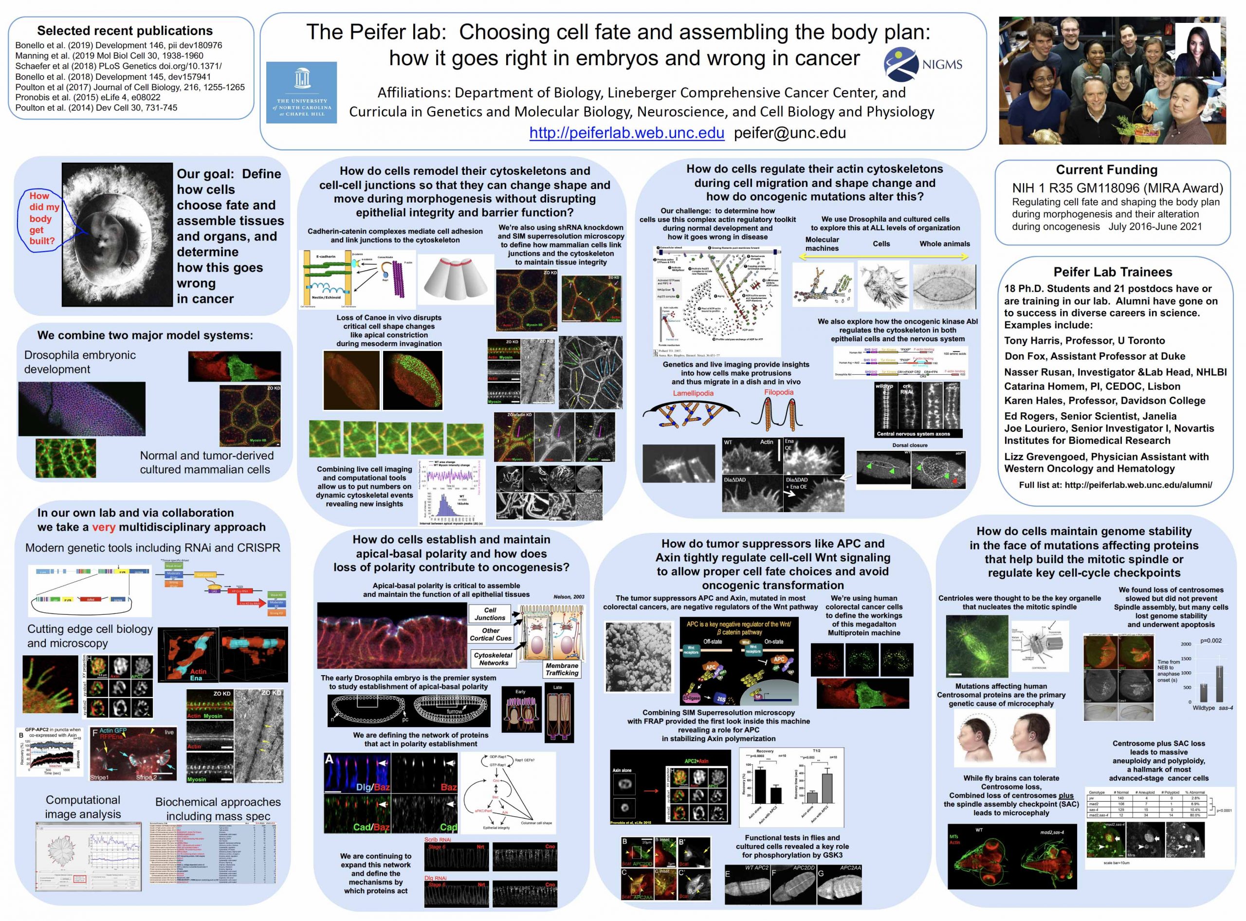
MORPHOGENESIS
We use developing embryos to explore the cell biology of morphogenesis—in this gastrulating embryo cytoskeletal (blue) and adhesion proteins (red) become reciprocally planar polarized.
-
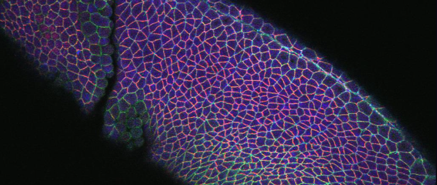
CELL CULTURES
We also use cultured Drosophila and mammalian cells to explore adhesion and Wnt signaling. This D17 cell illustrates how we can examine protrusive behavior in vitro.
-
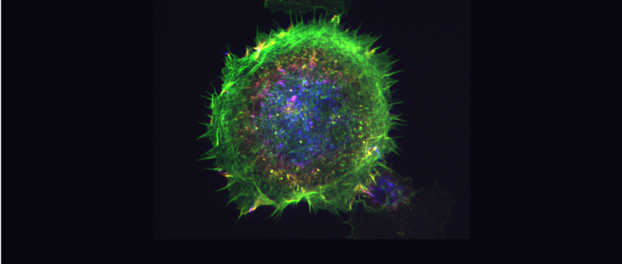
IN 3D
Our image analysis software allows us to explore protein localization in 3D. Here nuclei (green) collide during mitosis due to failure of the tumor suppressor APC to regulate centrosome (red) and cytoskeletal (blue) dynamics.
-
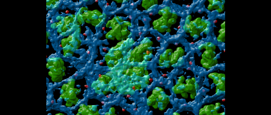
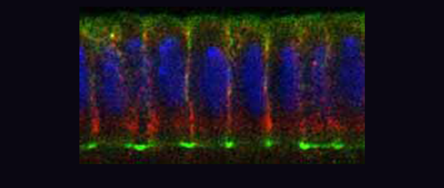
POLARIZATION
Drosophila embryos provide a superb place to assess the mechanisms by which cells polarize. This cellularizing embryo has basal actin rings (green) with basal junctions (red) just apical to them.
Lab Focus
WNT Signaling and APC
READ MORE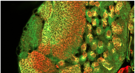
Wnt signals are one of the five signal transduction pathways that shape virtually all cell fates and which are inappropriately activated in most solid tumors. We explore novel biological roles for Wnt signaling during development, and seek to determine how the tumor suppressor APC regulates both Wnt signaling and the cytoskeleton.
Cell Adhesion and the Cytoskeleton
READ MORE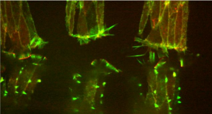
Our challenge is to alter the current static model of cell adhesion to explain the remarkable cellular events of morphogenesis that shape the embryonic body plan. To do so, we must understand the dynamic regulation of cell adhesion and the interactions between adhesion and the cytoskeleton.