Steph Nowotarski Alumna page
Background & Contact Information
Current: Postdoc with Alejandro Sánchez Alvarado, Stowers Institute/HHMI
University of North Carolina, Chapel Hill NC — Biology, 2006 -present
Fellowships and Awards: Graduate Fellowship, Cell and Molecular Biology Training Grant 2008-2009

Research Information
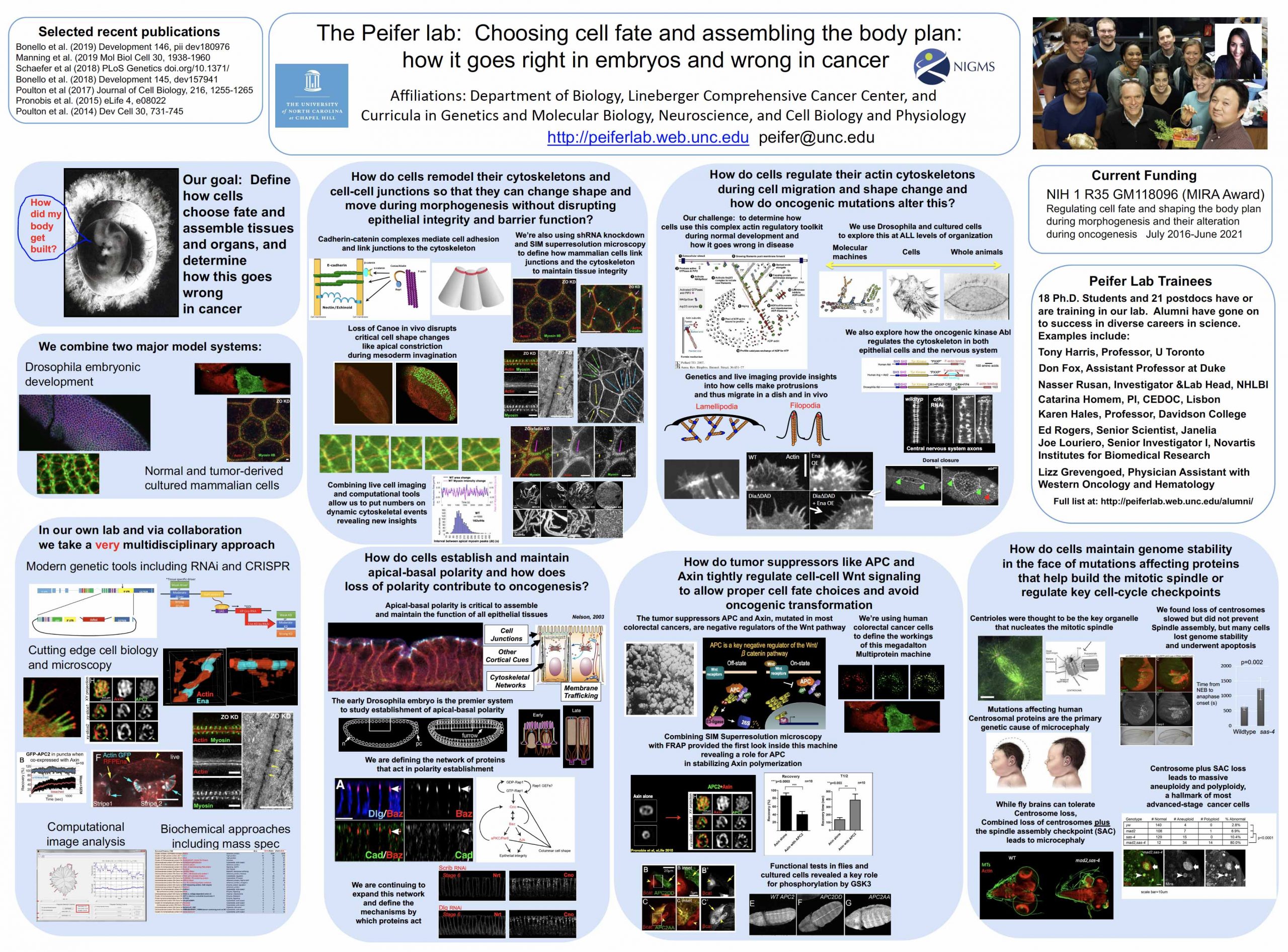
Rearrangement of the actin cytoskeleton mediates changes in cell shape, migration and subsequently cell behavior during morphogenesis, wound healing and homeostasis of adult tissue. This regulation is facilitated by an array of proteins that function in actin nucleation, polymerization, capping, bundling and severing. The decision to continue filament elongation or to terminate is a critical decision in the context of the cell, the tissue and ultimately the animal. Both the formin family protein Diaphanous (Dia), and the Enabled (Ena)/VASP family of proteins act at the barbed end to promote continued elongation, while Capping Protein (CP) functions antagonistically- acting at barbed ends to terminate elongation.
We previously analyzed the role of Ena during embryogenesis revealing that Ena plays a role in a subset of morphogenetic processes: germ band retraction, segment groove formation, head involution, and dorsal closure. We extended this analysis of Ena into oogenesis, finding it plays important roles in cortical integrity of germ-line nurse cells, and in formation of bundled actin filaments in nurse cells during the dumping process. Ena localizes to barbed ends of these specialized actin structures and appears to be negatively regulated by Ableson kinase in this context. We found CP is important for nurse cell dumping and plays a surprisingly critical role in oocyte specification. These data support an antagonistic relationship between Ena and CP in oogenesis. Current work focuses on further exploring the relationship between Ena and CP in the context of morphogenesis in collaboration with the Giniger Lab using both the nervous system and dorsal closure as models.
In addition to genetically interacting with CP, Ena also interacts with the formin Diaphanous ( see Colleen Guerin Bilancia’s page). This raises the question: Why have two actin elongation factors? We are currently dissecting the relationship between these two proteins in collaboration with the Kovar Lab. My continued work on this focuses on understanding how these proteins are regulated.
While the decision to continue or terminate a filament is important in the context of the cell, the differences between protrusive character between tissues is also important. We are currently exploring differential protrusive behavior between two types of cells in vivo. During dorsal closure lateral epidermal cells elongate along their dorsal ventral axis and migrate towards each other from opposing sides of the embryo. During this process the amnioserosa cells that sit between the lateral epidermal sheets constrict at their apices, are enclosed by the zippering epidermis, and are eventually eliminated. From previous and current work, it is clear that the leading edge cells of the epidermis along the amnioserosa/ epidermis boundary produce highly dynamic Ena based protrusions (yellow arrows above). However, both regulation of these LE filopodia and the mechanism leading to protrusions found in the amnioserosa cells is still unclear.
Publications
- Rogers, E.M., Spracklen, A.J., Bilancia, C.G., Sumigray, K.D., Allred, S.C., Nowotarski, S.N., Schaefer, K. N., Ritchie, B.J., and Peifer, M. (2016). Abelson kinase acts as a robust, multifunctional scaffold in regulating embryonic morphogenesis. Molecular Biology of the Cell 27, 2613-31.
- Nowotarski, S.H., McKeon, N., Moser, R.J., and Peifer, M. (2014). The actin regulators Enabled and Diaphanous direct distinct protrusive behaviors in different tissues during Drosophila development. Molecular Biology of the Cell 25, 3147-65.
- Kannan R, Kuzina I, WincovitchS, Nowotarski SH, Giniger E. (2014). The Abl/Enabled signaling pathway regulates Golgi architecture in Drosophila photoreceptor neurons. Molecular Biology of the Cell 25, 2993-3005.
- Nowotarski, S.H., and Peifer, M. (2014). Cell Biology: A tense but good day for actin at cell-cell junctions. Current Biology 24, R688-90.
- Bilancia, C.G. ,Winkelman, J.D., Tsygankov, D., Nowotarski, S.H. , Sees, J.A., Comber, K., Evans, I., Lakhani, V., Wood, W., Elston, T.C., and Peifer, M. 2014 . Enabled negatively regulates Diaphanous-driven actin dynamics in vitro and in vivo. Developmental Cell. In press.
- J-Y. Lin, W.J. Lin, W.-H. Hong, W.-C. Hung, S.H. Nowotarski, S. Montenegro Gouveia, I, Cristo and K.-H.Lin (2011) Morphology and organization of tissue cells in 3D microenvironment of monodisperse foam scaffolds. Soft Matter 7, 10010-10016.
- Gates J, Nowotarski SH, Yin H, Mahaffey JP, Bridges T, Herrera C, Homem CC, Janody F, Montell DJ, Peifer M. (2009) Enabled and Capping protein play important roles in shaping cell behavior during Drosophila oogenesis. Developmental Biology, Sep 1;333(1):90-107.
- Stevens T.L., Rogers E.M., Koontz L.M., Fox D.T., Homem C.C.F., Nowotarski S.H., Artabazon N.B., Peifer M. Using Bcr-Abl to Examine Mechanisms by Which Abl Kinase Regulates Morphogenesis in Drosophila. Molecular Biology o the Cell 2008 378 (19): 378-393.


