Colleen Bilancia Alumna page

Background & Contact Information
Currently: Cinical Cytogeneticist and Molecular Geneticist, Lineagan
ABMG Clinical Cytogenetics Fellow at Columbia University Medical Center (2014-2017)
Postdoc (2009 – 2014)
Education: Ph.D., Cell and Developmental Biology, University of Medicine and Dentistry of New Jersey and Rutgers University, Piscataway, NJ 2009
Embryology: Concepts and Techniques in Modern Developmental Biology, Marine Biological Laboratory, Woods Hole, MA 2006
B.A., Biology, La Salle University, Philadelphia, PA 2003
Fellowships & Awards: Postdoctoral Fellowship, UNC Developmental Biology Training Grant, 2009-2010
Email: colleen_guerin@yahoo.com
DOWNLOAD CV
Research Information
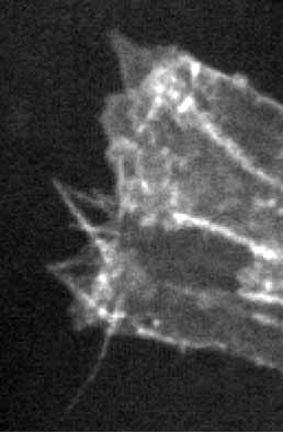
Cell protrusions are a hallmark feature of migrating cells during development and disease. During metastasis, cancer cells extend cell protrusions, become motile, and invade surrounding tissues. Cell protrusions are also required for cell migration during morphogenesis. As cells are specified during development, they must assume the correct position within the organism. Epithelial morphogenesis in the fruit fly, Drosophila melanogaster, provides an excellent model system for identifying regulators of cell protrusions. During dorsal closure, the sheet of epidermal cells migrates dorsally and encases the embryo. At the leading edge of migrating epidermal cells are highly dynamic cell protrusions. These protrusions are composed of both filopodia and lamellipodia, and previous studies in our lab discovered that a balance between these types of protrusions is necessary for proper morphogenesis. Two actin regulators, Diaphanous (Dia) and Enabled (Ena), are important modulators of cell protrusions at the leading edge during dorsal closure. Dia and Ena also play a role in cancer cell protrusions, which are required for metastasis. The roles of Dia and Ena in cancer protrusive behavior suggests cancer cells employ developmental signaling pathways and cytoskeletal machinery to drive migration and invasion. Therefore, understanding how Dia, Ena, and actin are regulated to promote cell protrusions during development will provide insight into how cancer cells acquire and regulate their migratory and invasive abilities.
Our preliminary studies found that Dia and Ena bind each other and regulate the type of cell protrusions formed by migrating epithelial cells in Drosophila. Therefore, I hypothesize that determining the nature and function of this interaction and identifying additional molecules that regulate Dia and Ena localization and activation will provide mechanistic insight into how cell protrusions are regulated. I will determine how Diaphanous and Enabled binding is mediated, how it affects actin dynamics at the filament level, and the role it plays in cell protrusions in vivo. I am also identifying regulators of Dia and Ena localization and activation to build a mechanistic view of cell protrusion formation.
Publications
- Rogers, E.M., Spracklen, A.J., Bilancia, C.G., Sumigray, K.D., Allred, S.C., Nowotarski, S.N., Schaefer, K. N., Ritchie, B.J., and Peifer, M. (2016). Abelson kinase acts as a robust, multifunctional scaffold in regulating embryonic morphogenesis. Molecular Biology of the Cell, in press.
- Bilancia, C.G. ,Winkelman, J.D., Tsygankov, D., Nowotarski, S.H. , Sees, J.A., Comber, K., Evans, I., Lakhani, V., Wood, W., Elston, T.C., and Peifer, M. 2014 . Enabled negatively regulates Diaphanous-driven actin dynamics in vitro and in vivo. Developmental Cell 28:394-408.
- Winkelman, J.D., Bilancia, C.G. Peifer, M., and Kovar, D.R.. 2014 . Ena/VASP Enabled is a highly processive actin polymerase tailored to self-assemble parallel-bundled F-actin networks with Fascin. Proceedings of the National Academy of Science USA 18;111(11):4121-6.
- Tsygankov, D., Bilancia, C.G., Vitriol, E.A., Hahn, K.M,, Peifer, M.*, Elston, T.C.* (*=co-corresponding). CellGeo: a computational platform for the analysis of shape changes in cells with complex geometries.. Journal of Cell Biology 204:443-60
- Guerin, C.M. and Kramer, S.G. (2009). RacGAP50C directs perinuclear γ-tubulin localization to organize the uniform microtubule array required for Drosophila myotube extension. Development, 136, 1411-1421.
- Guerin, C.M. and Kramer, S.G. (2009). Cytoskeleton Remodeling during Myotube Assembly and Guidance: Coordinating the Actin and Microtubule Networks. Commun Integr Biol. 2, 452-457.


